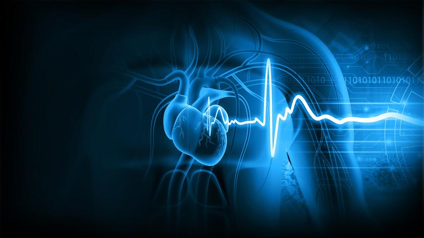
The human heart is not just a pump — it’s a beautifully timed orchestra. Every beat is a carefully coordinated cycle of events that ensures blood flows forward, oxygen gets delivered, and waste gets carried away. This repeating process is called the cardiac cycle, and it happens about 70–80 times per minute at rest, and much more when you’re running or stressed.
To really understand the cardiac cycle, let’s slow it down and walk through one single heartbeat — from the moment the atria fill to the moment the ventricles relax.
Step 1: Atrial filling and contraction (atrial systole)
The cycle begins with the atria (the two upper chambers) filling with blood. On the right side, blood comes from the body through the superior and inferior vena cava. On the left side, blood arrives from the lungs via the pulmonary veins.
As the atria fill, the valves between the atria and ventricles — the atrioventricular (AV) valves (tricuspid on the right, mitral on the left) — are open. Blood passively trickles down into the ventricles even before the atria contract.
Then, toward the end of atrial filling, the atria contract — a small “atrial kick” that tops up the ventricles with an extra 20–30% of blood.
- Key idea: Atrial systole is like the final push that ensures the ventricles are well loaded before they take on the big job of pumping blood out.
Step 2: Isovolumetric contraction
Now the ventricles are full. The atria relax, and the ventricles prepare to contract. As ventricular muscle fibers start to squeeze, pressure inside the ventricles rises quickly.
Here’s the critical moment: as soon as ventricular pressure exceeds atrial pressure, the AV valves snap shut. That’s the “lub” sound (S1) you hear with a stethoscope.
But at this point, the outlet valves (aortic and pulmonary) are still closed. So the ventricles are contracting, pressure is rising, but no blood is moving yet. This short phase is called isovolumetric contraction — “iso” means “same,” “volumetric” means volume. The blood volume inside the ventricle isn’t changing because all valves are shut.
- Key idea: This phase is like revving an engine before releasing the clutch — pressure builds, but nothing moves yet.
Step 3: Ventricular ejection
Once ventricular pressure becomes higher than the pressure in the aorta (on the left side) or pulmonary artery (on the right side), the outlet valves — the semilunar valves (aortic and pulmonary) — pop open.
Blood is then forcefully ejected:
- The right ventricle sends deoxygenated blood to the lungs through the pulmonary artery.
- The left ventricle pumps oxygenated blood into the aorta and throughout the body.
At first, ejection is rapid (rapid ejection phase). Then it slows down (reduced ejection phase) as ventricular contraction begins to wane.
This is the moment you feel as your pulse in the arteries.
- Key idea: Ventricular ejection is the “delivery phase” of the heartbeat — the real payoff for all the preparation.
Step 4: Isovolumetric relaxation
As soon as the ventricles finish ejecting most of their blood, pressure inside them begins to fall. Eventually, it drops below the pressure in the aorta and pulmonary artery. At this instant, the semilunar valves snap shut to prevent backflow. That closing makes the “dub” sound (S2) you hear with a stethoscope.
Now all valves are closed again — but this time, the ventricles are relaxing, not contracting. Volume remains the same for a short period because no blood enters or leaves. This is isovolumetric relaxation.
- Key idea: This is like a pressure release phase — the ventricles are relaxing but not yet receiving blood.
Step 5: Ventricular filling
When ventricular pressure falls below atrial pressure, the AV valves open again. Blood that has been accumulating in the atria now rushes into the ventricles. This happens in two stages:
- Rapid filling: blood flows quickly into the ventricles due to the pressure gradient.
- Slow filling (diastasis): as pressures equalize, filling slows down until the next atrial contraction tops things up again.
And just like that, we’re back to Step 1 — atrial systole — and the cycle repeats.
- Key idea: Ventricular filling is the “reset” step, ensuring the ventricles are primed for the next beat.
The Wiggers diagram: tying it all together
Medical students are often shown the Wiggers diagram — a graph that combines pressure, volume, heart sounds, and ECG waves. It looks intimidating, but it’s just the story above drawn out in curves.
- The P wave on ECG corresponds to atrial contraction.
- The QRS complex signals ventricular depolarization (leading to contraction).
- The T wave marks ventricular repolarization (relaxation).
When you map these onto valve movements and heart sounds, the puzzle pieces click.
Why this cycle matters clinically
- Heart sounds:
- S1 = closure of AV valves at start of ventricular systole.
- S2 = closure of semilunar valves at start of ventricular diastole.
- Extra sounds (S3, S4) often indicate pathology (like heart failure).
- Pulse pressure & stroke volume:
- The ventricular ejection phase determines how much blood (stroke volume) gets pumped out. Problems here lead to weak or absent pulses.
- Valve diseases:
- Stenosis (narrowing) or regurgitation (leakage) alter the normal sequence. For example, mitral regurgitation means AV valves don’t close properly during systole, so blood leaks backward.
- Shock and heart failure:
- In cardiogenic shock, the ventricle fails to generate enough pressure in systole.
- In diastolic heart failure, the filling phase is impaired, so preload is reduced.
Everyday analogies to cement the cycle
- Think of the heart like a two-cylinder engine with intake (filling), compression (isovolumetric contraction), power stroke (ejection), and exhaust (relaxation).
- Or picture it as a washing machine: water fills (atrial/ventricular filling), lid locks (valves close), spin cycle (ejection), pause (relaxation), then repeat.
Final recap (simple version)
- Atria contract → ventricles topped up.
- Ventricles start to contract → AV valves close (S1).
- Pressure builds with all valves shut → isovolumetric contraction.
- Semilunar valves open → blood ejected.
- Semilunar valves close (S2) → isovolumetric relaxation.
- AV valves reopen → ventricles fill again.
One heartbeat. Repeat about 100,000 times per day.


