🔹 What is an Anastomosis?
In medicine, anastomosis means a connection or joining between two tubular structures. In the cardiovascular system, it usually refers to the joining of blood vessels that allows alternate routes for blood flow.
So, cardiopulmonary anastomosis = the vascular connections between the heart and the lungs that ensure continuous blood supply and oxygen exchange, even if one route is blocked or compromised.
🔹 Anatomy of Cardiopulmonary Anastomoses
The lungs and heart are richly supplied by two circulatory systems:
- Pulmonary circulation – carries deoxygenated blood from the right ventricle to the lungs for oxygenation.
- Bronchial circulation – part of systemic circulation; arises from the thoracic aorta and supplies the bronchi, connective tissue, and pleura of the lungs.
At the microscopic level, these two systems interconnect through tiny vascular anastomoses.
- Bronchopulmonary anastomoses → connections between bronchial arteries/veins and pulmonary arteries/veins.
- They exist mainly at the level of respiratory bronchioles and alveoli, where small bronchial vessels blend with pulmonary capillaries.
- This creates a “safety net” circulation for the lungs.
🔹 Functional Importance
Why does this matter?
- Backup oxygen supply – If pulmonary circulation is compromised (e.g., a small embolus), the bronchial circulation can still deliver oxygen and nutrients to lung tissue.
- Pressure equalization – Anastomoses help balance pressure differences between systemic bronchial vessels and low-pressure pulmonary vessels.
- Gas exchange support – They maintain perfusion around alveoli to optimize oxygen exchange.
- Pathophysiology – In diseases, these anastomoses can become exaggerated or dysfunctional.
🔹 Physiological Mechanisms
- The pulmonary arteries bring large volumes of blood at low pressure for oxygen exchange.
- The bronchial arteries (from systemic circulation) bring smaller amounts of blood at higher pressure to nourish the lung tissue.
- The anastomoses act like “bridges” between these two systems, allowing blood to move if one side is blocked or overloaded.
A classic example:
- If pulmonary arteries are obstructed, blood may “shunt” from bronchial arteries → pulmonary capillaries.
- If bronchial veins are congested, some blood can drain into pulmonary veins (this partly explains why a small fraction of bronchial blood contributes to left atrial oxygenated blood).
🔹 Biochemistry Angle
At the tissue level:
- Oxygen demand is met both by direct oxygen in inspired air and by vascular supply.
- Bronchial circulation delivers glucose, nutrients, and oxygen to airway cells, while pulmonary circulation primarily ensures gas exchange at alveoli.
- Anastomoses ensure both processes overlap smoothly.
🔹 Clinical Relevance
- Pulmonary Embolism (PE):
Small emboli may not cause infarction because bronchial circulation sustains the affected tissue via anastomoses. Large emboli, however, can overwhelm both systems → infarction. - Chronic Lung Diseases (like COPD):
These conditions often increase bronchial-to-pulmonary anastomoses, contributing to abnormal blood flow and sometimes hypoxemia. - Congenital Heart Disease:
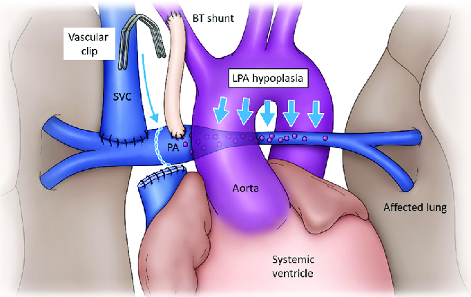
Abnormal cardiopulmonary anastomoses can contribute to right-to-left or left-to-right shunts. - Surgery/Transplant:
In lung transplants, surgeons must preserve bronchial circulation; otherwise, ischemia may occur in airway tissues despite normal pulmonary flow.
🔹 Summary for Students
Think of cardiopulmonary anastomosis as the network of backup bridges between systemic bronchial vessels and pulmonary vessels.
- Anatomy: Bronchial ↔ Pulmonary connections
- Function: Safety net, oxygen supply, pressure balance
- Biochemistry: Supplies nutrients and oxygen to lung tissues
- Clinical importance: Explains survival of lung tissue in small emboli, changes in chronic disease, and surgical considerations
👉 If the pulmonary highway is blocked, bronchial side roads can still keep traffic moving.
Mnemonic to Remember:
“Bronchial feeds, Pulmonary exchanges, Anastomosis connects.”


