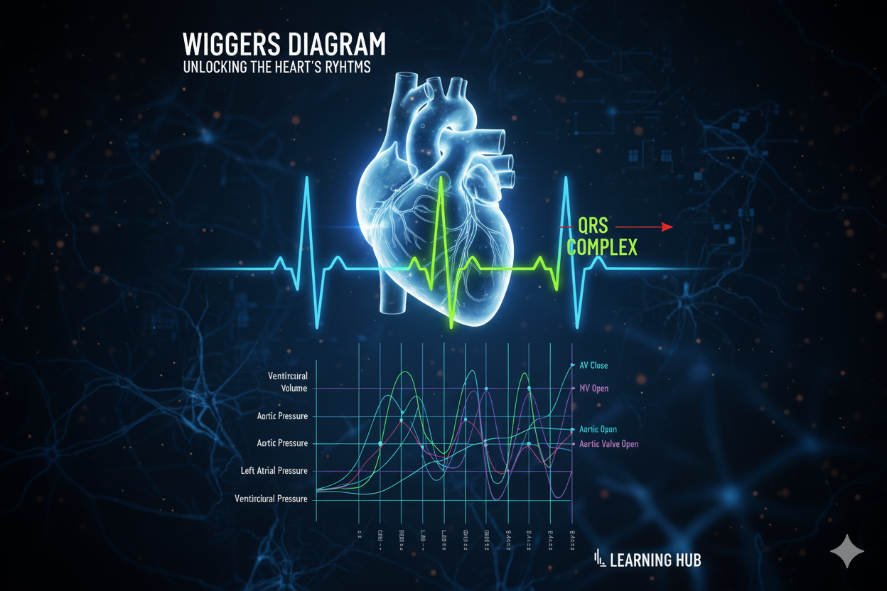
If you’ve ever opened your physiology textbook and seen the Wiggers Diagram, you probably thought: “This looks like a spaghetti chart!”
Don’t worry. By the end of this guide, you’ll understand exactly what’s going on. The Wiggers Diagram is simply a timeline of the cardiac cycle—showing how pressure, volume, ECG, and heart sounds all happen together in the heart.
Let’s break it down.
What is the Wiggers Diagram?
The Wiggers Diagram is a classic graph in physiology that represents all the events of the cardiac cycle in one picture.
It combines:
- Electrical activity (ECG)
- Pressure changes (atria, ventricles, aorta)
- Volume changes (in the ventricles)
- Heart sounds (phonocardiogram: S1, S2, etc.)
This is why it looks so complicated—because everything is happening at once. But if we follow it layer by layer, it makes sense.
The Players (What the Graph Tracks)
Before we trace the cycle, let’s know what’s on the diagram:
- ECG (Electrical activity) – tells us when the atria or ventricles depolarize.
- Atrial pressure curve – small wave changes when atria contract or relax.
- Ventricular pressure curve – sharp rise during systole, drop during diastole.
- Aortic pressure curve – reflects blood being pushed into circulation.
- Ventricular volume curve – how much blood fills/empties the ventricle.
- Heart sounds (S1, S2) – valve closures.
Step-by-Step Through the Wiggers Diagram
Think of the cycle as phases. One heartbeat (≈0.8 sec) includes:
Phase 1: Atrial Systole (Atrial contraction)
- Trigger: P wave on ECG (atrial depolarization).
- What happens: atria contract and push the last bit of blood into ventricles (the “atrial kick”).
- Pressure: atrial pressure rises slightly.
- Volume: ventricular volume increases (end-diastolic volume reached).
- Sound: no major heart sound here (but sometimes S4 in pathology).
💡 Key idea: Atrial contraction “tops off” ventricular filling.
Phase 2: Isovolumetric Contraction
- Trigger: QRS complex (ventricular depolarization).
- What happens: ventricles contract but all valves are closed.
- Pressure: ventricular pressure shoots up rapidly.
- Volume: no change (iso-volumetric = same volume).
- Sound: S1 (first heart sound) → closure of AV valves (mitral + tricuspid).
💡 Key idea: Pressure rises without blood leaving the ventricle.
Phase 3: Ventricular Ejection
- What happens: once ventricular pressure exceeds aortic pressure, the aortic valve opens.
- Pressure: ventricular and aortic pressures both rise.
- Volume: ventricular volume drops as blood is ejected.
- ECG: ST segment → T wave begins (ventricular repolarization).
- Sound: none (but sometimes Systolic murmur in disease).
💡 Key idea: Blood is pumped into systemic circulation here.
Phase 4: Isovolumetric Relaxation
- Trigger: End of T wave (ventricular repolarization).
- What happens: ventricles relax; all valves are closed.
- Pressure: ventricular pressure falls rapidly.
- Volume: no change.
- Sound: S2 (second heart sound) → closure of semilunar valves (aortic + pulmonary).
💡 Key idea: The heart relaxes, pressure falls, but no blood enters yet.
Phase 5: Ventricular Filling (Diastole)
This has two parts:
- Rapid filling: AV valves open → blood flows quickly from atria to ventricles.
- Ventricular volume rises sharply.
- Sometimes produces S3 sound (normal in children, abnormal in adults).
- Slow filling (diastasis): flow slows down until the next atrial contraction.
💡 Key idea: Ventricles fill passively, getting ready for the next atrial systole.
Linking Everything Together
Now let’s align all the events in one heartbeat:
- ECG → P wave = atrial contraction.
- QRS complex = ventricular contraction starts → S1 (AV valves close).
- Ventricular pressure rises → aortic valve opens → blood ejected.
- T wave = ventricles repolarize → S2 (aortic valve closes).
- Ventricular relaxation → AV valves open → filling phase begins again.
The cycle repeats every 0.8 seconds (at 75 bpm).
Why the Wiggers Diagram Matters
- Clinical relevance:
- Helps explain murmurs (e.g., systolic vs diastolic).
- Links ECG findings to mechanical events.
- Useful for interpreting hemodynamic tracings in ICU or cardiology.
- Physiology relevance:
- Connects electrical, pressure, volume, and sound events in one frame.
- Shows how tightly structure and function are linked in the heart.
Take-Home Points
- The Wiggers Diagram is a timeline of the cardiac cycle.
- It tracks ECG, pressure, volume, and sounds together.
- Break it into phases: atrial systole → isovolumetric contraction → ejection → isovolumetric relaxation → filling.
- Heart sounds: S1 = AV valves close, S2 = semilunar valves close.
- Once you see the pattern, the “spaghetti” makes perfect sense.